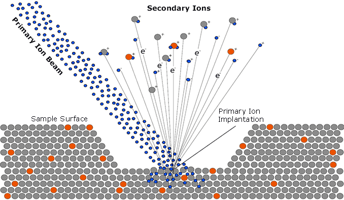
Time-of-Flight Secondary Ion Mass Spectrometry (ToF-SIMS) is an analytical method where the sample surface is bombarded with a pulsed primary ion beam. The primary ion energy is transferred to the target atoms via a series of collisions; a process known as the “collision cascade.” The collision cascade transports a small part of the energy back to the surface resulting in the desorption or sputtering of electrons, neutrals and secondary ions. The secondary ions aretransferred into the mass spectrometer and their mass is determined by measuring the flight time to the detector. Thus, the chemical composition of the surface can be probed with high sensitivity. The technique provides information on the atomic and the molecular composition of the uppermost 1 – 3 monolayers with sensitives at the ppm level. By combining imaging and depth profiling, ToF SIMS analysis enables the characterization of the 3D chemical composition of a sample.
In spectrometry mode a total mass spectrum of the surface region is acquired. These spectra are usually recorded with high mass resolution and use a low number of primary ions. The high mass resolution is necessary for reliable identification of secondary ion signals and corresponding chemical assignment. The limited number of primary ions guarantees that the detected signals are representative of the original chemical composition of the sample surface.
For the acquisition of mass resolved secondary ion images, a focused primary ion beam is used to probe the surface of interest. A complete mass spectrum is recorded for each pixel. The intensities of secondary ions are color coded resulting in a mass resolved intensity map of the lateral distribution of secondary ion emission. Fields of view of up to 500 x 500 μm2 can also be analyzed by rastering the primary ion beam.Larger areas (500 x 500 µm2 … 9 x 9 cm2) can be analyzed using a stage scan raster. The lateral resolution of the secondary ion images ranges from 300 nm to 2 µm depending upon the specific ToF SIMS Analysis conditions.
Compositional depth profiling is also possible in order to investigate the chemical composition of a sample as a function of depth. In ToF SIMS Analysis, two different ion beams are used for data acquisition: A sputter beam (e.g. O2+, Cs+, C60, or Ar GCIB) is applied to erode the sample while a second ion beam (analysis beam e.g. Bix+) is used to chemically characterize the resulting crater bottom. Typically, O2+or Cs+ depth profiles only show the distribution of elements because the massive sample erosion causes a destruction of molecular structures. However, when a Arn+ gas cluster ion beam (GCIB) is used as the sputter beam, molecular depth profiling is possible. The low energy per atom of the Arn+ GCIB extends the “static” limit, maintaining organic information under sputtering. With ToF-SIMS depth profiling, 3D visualization of complex sample structures is possible by combining spectral, imaging and depth information.


